- Objective and subjective (quantitative) Algesimetry
An advanced approach in pain and analgesia research is the human experimental model utilizing the objective-quantitative, high-resolution laser algesimetry. This method is a well-established and validated alternative to the pure subjective pain estimations of patients in clinical conditions of pain management (e.g. working with visual analogue scales, VAS, categorical scales or pain diaries). A CO2-laser is used to initiate Laser (radiant-heat) evoked potentials (LEP) by selective Aδ- and C-fibre stimulation (incl. TRPV1-3 receptors), which are recorded in the electroencephalogram (EEG) from the Vertex scalp position (see basic recording and online evaluation at Laser Principle). The anti-nociceptive/anti-hyperalgesic properties of analgesic drugs and topicals can be demonstrated objectively and quantitatively by alterations in LEP variables (primarily reductions of amplitudes) e.g. versus placebo or a reference drug/ analgesic.
The major advantage of this thermo-nociceptive stimulation with a CO2-laser is, that heat-sensitive ionic channels of the polymodal nociceptors of Aδ- (thinly-myelinated) and C-fibre (non-myelinated) types are selectively stimulated – due to the exact skin layer penetration of CO2-laser radiation to free nociceptor terminals without any direct skin contact. The radiation of this type of laser is independent of the melanin and haemoglobin content of skin due to the spectral emission range of these laser devices in the far infrared (important for work in inflamed and pigmented skin). The resulting two main components of the 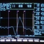 EP (N2 and P2, see figure left) are evaluated with regard to their complex peak-to-peak (PtP) amplitude, as well as with regard to the single N2-component, which mainly reflects ‘peripheral’ nociceptive processing in central mechanisms/ structures, and the single P2-component, which mainly reflects ‘central’ nociceptive processing in central mechanisms/ structures. A main feature of the laser stimulation is that there is no underlying and resulting placebo effect by the application itself. Laser nociceptive stimuli can be applied to normal or sensitized skin in a disease-like/ “healthy patient” manner (e.g. to UVB-irradiated/ inflamed/ ”sun-burned” skin, the latter being a “real disease”, including all the important parts of the developing, mediator-induced inflammatory (e.g. cyclo-oxygenase) cascade as seen in clinical conditions, like acute post-traumatic/ post-operative pain and hyperalgesia, as well as to capsaicin-irritated skin, with developing neurogenic, mixed primary and secondary hyperalgesia and allodynia – induced by an ongoing nociceptive, spinal input.
A considerable number of typical and atypical analgesic compounds, respectively compound classes and topicals, have been investigated at HPR over the years (see Database analgesics LSEP_HPR) and may help to reclassify newer entities with regard to their general efficacy, their onset of efficacy as well as to their time- and dose-efficacy.
Find out more about Algesimetry…
- Inflammatory measurements (intensity, colour)
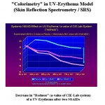 The “redness” of the UVB– and capsaicin-induced skin erythema and its time course can be quantified by objective skin reflection spectrometry (SRS, inflammatory and neurogenic erythema intensity measurement – using the a-value of the Lab-system according to the CIE-definition or by quantitative colour spectra; see figure left with fading of “steady-state” UV-erythema under two different NSAID doses). The size of the capsaicin-induced flare area can be determined by computer planimetry. SRS can also be used for the measurement of hematoma intensities (e.g. after own-blood-injections and their timely colour fading with and without topical treatment).
Find out more about Inflammatory measurements…
- Evoked potentials:
Standardised visual, auditory, or somatosensory signals/ stimuli are applied over the main assessment days during a trial period. Before and after the actual administration of medications event related/ evoked potentials (ERPs, EPs) are recorded e.g. from Vertex- 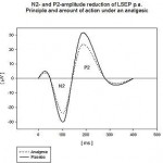 and Occipital-EEG positions (AEPs, SEPs, VEPs) and evaluated with regard to their amplitudes and latencies (see figure left) as an objective-quantitative measure of so-called “specific” vigilance- and nociceptive-related CNS-responses (specific afferent pathways of peripheral and CNS information processing). These methods are subject to no or only to minor habituation, when certain prerequisites are taken into account – e.g. as random stimulus presentation. The attention of the subjects is kept stable during all Evoked Potential procedures by performing a simultaneous pursuit tracking task (PTT, target-pursuer paradigm). A further distraction from external disturbances will be done by presenting binaural “white” noise (with a sound pressure of 80 to 90 dbA, the latter not in case of auditory evoked potentials, AEPs).
Find out more about Evoked potentials…
- Standard-/ provocation-EEG and EEG-brain mapping:
The resting, vigilance-controlled and task-related standard EEG is recorded and its absolute and relative spectral power as well as its dominant frequencies are analysed for the evaluation of “non-specific” vigilance states of CNS (spontaneous endogenous- or exogenous-induced fluctuations of vigilance), e.g. under drug or hypoxic influence. Provocation methods, as photic stimulation/ driving, hyperventilation and complete sleep deprivation, can be applied to detect epileptic/ hyper-excitatory tendencies in CNS of subjects (spike and wave [s/w] detection). Up to 32 EEG-channels can be evaluated simultaneously.
Find out more about EEG and EEG-brain mapping…
- Oculodynamic test (ODT):
Saccadic eye movements to (5) fixed positions with (5) randomly presented types of targets 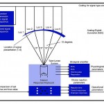 in the horizontal visual axis (for principle see figure left) and following a fixed timely sequence (1s) are recorded by surface electrodes on both eyes. After automatic online artefact-rejection the latency (in ms, a parameter equivalent to polysynaptic retino-ponto-muscular reflex time), the angular velocity (in degrees/s, a parameter mirroring the efferent and myogenic outcome at the eye-muscles – the latter displaying the highest innervation density of all muscular structures in the human body) and the time to fixation (in ms, a parameter representing retinal, neuronal and central components) of each eye movement/ saccade at the left and right eye are evaluated (600 to 1200 per session). These objective-dynamic parameters of vigilance, psychomotor performance and myogenic conditions are not influenced by motivation and learning and are not subject to habituation. During the ODT a complex choice reaction task (CRT, see figure above) is run simultaneously. The combination of both delivers a highly complex and objective-quantitative image of stimulative and sedative, as well as myogenic, drug influences on specific afferent and efferent CNS processing, which is also linked to every-day activities e.g. like car-driving-ability, piloting and handling machinery.
- Complex choice reaction task (CRT):
This test is normally integrated in the saccadic ODT procedure (see figure above), but can also be used – as a “stand-alone” test. The subject uses an ergometrically designed key board for the identification and coding of 5 different light signals (modified Pflüger’s hooks) in the horizontal visual axis (saccades of 15, 30, 45 and 60°). The mean number of correctly and incorrectly identified light signals (per min) reflects the quality of attentional/ cognitive information processing in CNS-performance. The mean complex choice reaction time (ms) is a speed-oriented measure of the psychomotor performance (afferent and efferent pathways of CNS-processing). Both parameters are closely attached to the necessary daily activities – as mentioned above.
- Electroretinogram (ERG):
The ERG is a special type of evoked potentials (run at the eyes). HPR has developed a very comfortable method of recording the ERG with surface skin electrodes. The retina is the only extra-cranial organ which contains functional glia cells (the “Müller Cells”) .The ATP-dependent K-pump system of the retina is the basis of the most important electrophysiological retinal signal of the eye – the b-wave. As the retina (and thus the ERG) is extremely sensitive to hypoxia, the ERG can be used to detect drug effects which restore hypoxia-induced functional deficits by either affecting the local concentrations of neurotransmitters and/or the sensitivity/ number of these receptors, or by influencing the local and/or the general CNS metabolism by improving the supply with essential substrates (as O2, glucose, ATP) and transmitters. During the ERG sessions a simultaneous measurement of the concomitant visual evoked potential (VEP) response can be recorded and evaluated (at occipital cortex). ERG also has shown changes in “membrane-active” compounds, e.g. as seen after multiple dosing of hypolepemics (in healthy volunteers!).
- Pursuit tracking test (PTT)/Eye-Hand-Coordination:
This vigilance/ psychomotor performance testing situation demands the subject to 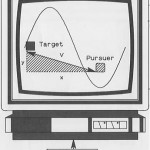 follow and cover a randomly moving target on a computer screen with a pursuer signal by means of a joy stick. The mean “error vectors” per minute (v from x- and y-axis deviation, see figure left) between target and pursuer will be permanently evaluated. They reveal information on the steering/ coordination capabilities under alertness and fatigue – as well as under drug conditions, needed in car driving, piloting and handling complicated machinery (eye-hand coordination/ visu-motor capability). The tracking procedure is also used in the diverse EP-sessions (AEP, LEP, SEP, VEP) to stabilize the general vigilance conditions of the subjects – as well as to distract them from ongoing measurement procedures.
- Evaluation of CRF-data and p&p-questionnaires with computer-assisted methods:
Fill-ins of CRFs, multiple choice questionnaires (e.g. psychiatric scales), visual analogue scales and coded experimental and clinical pd-parameters (BP, HR, temperature etc.) can be automatically evaluated and stored in a “protected and sealed mode” with an individual checksum by aid of computers (e.g. in Excel sheets for later data management and statistical procedures).
- Visual analogue scales (100mm):
Instead of using uni- and/or bipolar 100 mm paper and pencil (p&p) visual analogue scales HPR developed a direct VAS fill-in mode on tablet-PC, which is used for the subjective online rating/ scoring of diverse personal status conditions/ polarities, e.g. items like sedation/excitation, anxiety/calmness, pain/analgesia and other relevant parameters to be scored (data are directly transcribed into “protected” Excel sheets).
Find out more about Visual analogue scales via Algesimetry and Visual analogue scales via CNS performance…